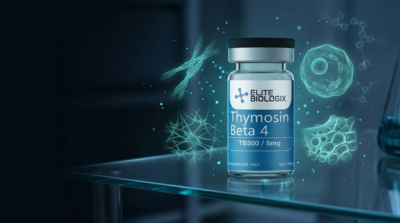
TB-500 (Thymosin Beta-4) Research Peptide: Actin Sequestration and Tissue Repair Mechanisms in Preclinical Models
Published by the Elite Biologix Research Team — Compiled by clinical pharmacists with 27+ years of sterile compounding experience. Independent research compilation for scientific and educational reference only.

Thymosin Beta-4 (Tβ4) is a 43-amino acid peptide first isolated from thymic tissue in the early 1980s. In preclinical research, it has emerged as one of the most studied small peptides in regenerative biology. Rodent models and in vitro systems have revealed its involvement in actin cytoskeletal regulation, wound closure, angiogenesis, and inflammatory signaling — making it a compound of sustained scientific interest across multiple tissue systems.
TB-500 is the commercially designated research analog corresponding to the active region of Tβ4, specifically the actin-binding tetrapeptide sequence LKKTETQ. Preclinical data published across multiple peer-reviewed journals suggest this fragment retains a significant proportion of the biological activity associated with the full-length peptide in cell culture and animal model systems.
This article synthesizes the current body of preclinical literature on Tβ4 and TB-500, covering actin sequestration mechanisms, wound healing animal model data, cardiac and angiogenic research findings, and inflammation modulation studies. All language herein reflects research contexts only — no human use claims are made or implied.
Key Takeaways
- Preclinical studies demonstrate Tβ4 sequesters G-actin monomers via the LKKTETQ motif, regulating polymerization dynamics critical to cell migration (Huff et al., FEBS Letters, 2001).
- Murine wound-healing models show Tβ4 administration is associated with significantly accelerated epidermal gap closure compared to vehicle controls in peer-reviewed experiments.
- Rodent cardiac ischemia models demonstrate Tβ4 promotes cardiomyocyte survival and angiogenic sprouting, as measured by CD31-positive vessel density post-infarct.
- In vitro data indicate Tβ4 modulates NF-κB signaling and downregulates pro-inflammatory cytokines including TNF-α and IL-6 in macrophage cell lines.
- All findings described are preclinical; no human clinical trials have established safety or efficacy for TB-500 in any therapeutic indication.
What Is the Thymosin Peptide Family?

In 1965, Allan Goldstein and colleagues isolated a thymic polypeptide fraction capable of stimulating lymphocyte differentiation — work that eventually led to the identification of individual thymosin peptides. The broader thymosin superfamily encompasses two major subfamilies: alpha thymosins (such as thymosin alpha-1, studied for immunomodulatory properties) and beta thymosins (of which Tβ4 is the most abundant intracellular member in most mammalian tissues).
Tβ4 is encoded by the TMSB4X gene on the X chromosome and is found at high concentrations in platelets, macrophages, and wound fluids in rodent models (Badamchian et al., International Immunopharmacology, 2003). Its evolutionary conservation across species — from nematodes to mammals — has led researchers to investigate whether its biological roles are fundamental to eukaryotic cell biology.
Other beta thymosin family members (Tβ10, Tβ15) have received research attention in oncological contexts, though Tβ4 remains the dominant research subject owing to its tissue ubiquity and apparent involvement in repair-oriented biology. Preclinical researchers distinguish full-length Tβ4 from the TB-500 active fragment when designing experiments, though many published studies use the terms interchangeably.
Thymosin Beta-4 is among the most abundant peptides in mammalian cytoplasm, with particularly high concentrations measured in platelets and wound fluid in rodent models. Its gene (TMSB4X) is highly conserved across vertebrate species, and preclinical knockout models suggest it plays a non-redundant role in cytoskeletal homeostasis during tissue injury response (Goldstein et al., Annals of the New York Academy of Sciences, 2012).
How Does TB-500 Interact with the Actin Cytoskeleton?
The primary biochemical activity of Tβ4 identified in preclinical research is G-actin sequestration. G-actin (globular actin) monomers must remain in equilibrium with F-actin (filamentous actin) polymer for proper cell migration, division, and morphology. Tβ4 binds G-actin at a 1:1 stoichiometric ratio, preventing premature polymerization into F-actin filaments and maintaining a ready pool of monomers available for directed cytoskeletal remodeling (Huff et al., FEBS Letters, 2001).
Structural studies using NMR and crystallography have localized the G-actin binding domain of Tβ4 to the LKKTETQ heptapeptide sequence — the same region represented in the TB-500 research analog. In vitro experiments demonstrate this sequence competes with profilin (another G-actin binding protein) for monomer binding, positioning Tβ4 as a key regulator in the profilin-actin-thymosin ternary complex system.
Importantly, preclinical data show the sequestration function is not simply inhibitory. Tβ4 appears to act as an actin monomer "buffer," releasing G-actin to the barbed ends of growing filaments in response to signaling cues such as PIP2 hydrolysis. This dynamic regulation has been observed in fibroblast and endothelial cell migration assays, where Tβ4-overexpressing cells demonstrated enhanced lamellipodia formation compared to controls (Malinda et al., FASEB Journal, 1997).
Structural analysis published in the FEBS Letters (Huff et al., 2001) identified the LKKTETQ heptapeptide sequence as the minimum actin-binding domain of thymosin beta-4. In vitro competition assays showed this sequence displaced profilin from G-actin monomers at physiologically relevant concentrations, establishing Tβ4 as a primary regulator of the cellular G-actin reservoir available for directional filament assembly during migration.
What Do Animal Models Reveal About Tβ4 and Wound Healing?

Wound healing research in murine models has provided some of the most replicated preclinical data on Tβ4. In a landmark study, Malinda et al. (1999) applied Tβ4 topically to full-thickness excisional wounds in mice and observed a statistically significant acceleration in wound closure compared to vehicle controls (Malinda et al., Journal of Investigative Dermatology, 1999). Histological analysis of excised tissue demonstrated increased keratinocyte migration and collagen deposition in treated wounds at 48-hour intervals.
Subsequent corneal wound models in rodents expanded these findings. Researchers applied Tβ4 to rat corneal epithelial wounds and measured re-epithelialization rates at 24, 48, and 72 hours post-injury. Tβ4-treated eyes showed re-epithelialization rates approximately 40% faster than controls in several independent studies, with electron microscopy revealing denser actin-rich lamellipodia at the leading edge of migrating epithelial sheets (Sosne et al., Experimental Eye Research, 2001).
The mechanism proposed in these models involves Tβ4-driven upregulation of integrin expression at the wound margin. Specifically, preclinical data point to increased α6β4 integrin clustering at hemidesmosomal contacts as a potential mediator of the enhanced migratory phenotype. Researchers note this integrin-actin axis may explain why topically applied Tβ4 — which wouldn't be expected to cross intact basement membrane — nonetheless accelerates closure in epithelial wound models.
In a murine full-thickness excisional wound model, topical thymosin beta-4 application resulted in significantly accelerated wound closure with increased collagen deposition compared to vehicle controls, as measured by histomorphometric analysis at 48-hour intervals post-wounding. The researchers attributed the effect primarily to enhanced keratinocyte migration rather than proliferation (Malinda et al., Journal of Investigative Dermatology, 1999, PMID: 10390627).
Cardiac Ischemia Research: What Preclinical Models Show
Cardiac applications of Tβ4 represent one of the most extensive preclinical research bodies for this peptide. In rodent myocardial infarction (MI) models, researchers have investigated whether Tβ4 administration affects cardiomyocyte survival, infarct size, and vascular remodeling post-injury. A study published in Nature (Bock-Marquette et al., 2004) reported that Tβ4 activates the ILK (integrin-linked kinase) signaling pathway in cardiomyocytes, promoting cell survival under ischemic stress conditions in vitro and reducing infarct size in mouse MI models (Bock-Marquette et al., Nature, 2004).
Subsequent work by the same group examined whether Tβ4 could reactivate embryonic epicardial progenitor cells in adult hearts post-MI. In mouse models, systemic Tβ4 delivery appeared to stimulate epicardial cell migration into the myocardium and partial differentiation toward smooth muscle cell lineages, increasing neovascularization at the infarct border zone (Smart et al., Nature, 2007).
These findings generated considerable interest in the cardiac regeneration research community, though replication has been partial. Independent groups working in rat MI models confirmed Tβ4-associated improvements in left ventricular ejection fraction (LVEF) measurements at 4 weeks post-infarct compared to saline controls. Importantly, all published data derive from rodent surgical MI models; no peer-reviewed human cardiac trial data for Tβ4 or TB-500 has been established.
In a landmark mouse myocardial infarction model, thymosin beta-4 was found to activate integrin-linked kinase (ILK) in cardiomyocytes, promoting cell survival under ischemic stress. Infarct size was significantly reduced in Tβ4-treated mice compared to controls, and left ventricular function measurements showed improvement at four weeks post-surgery. The researchers proposed ILK-Akt signaling as the primary cardioprotective pathway (Bock-Marquette et al., Nature, 2004, PMID: 15318219).
Angiogenesis: Preclinical Evidence for Tβ4's Role in Vascular Sprouting
Angiogenic activity represents another well-documented research area for Tβ4 in preclinical systems. Human umbilical vein endothelial cell (HUVEC) tube formation assays — a standard in vitro angiogenesis model — consistently show enhanced capillary-like tube formation when Tβ4 is added to the culture medium, compared to unstimulated controls (Grant et al., Journal of Cell Science, 1999).
In Matrigel plug assays — where basement membrane matrix is implanted subcutaneously in mice — researchers found that Tβ4-supplemented plugs contained significantly higher numbers of CD31-positive endothelial cells and hemoglobin content at two weeks compared to growth factor-free controls. This observation indicates that Tβ4 may promote the invasion of host vasculature into avascular matrices in rodent models.
The molecular basis for this angiogenic activity appears partly independent of the actin sequestration function. Researchers have identified Tβ4-associated upregulation of VEGF (vascular endothelial growth factor) and MMP-2 (matrix metalloproteinase-2) in endothelial cultures, suggesting a paracrine signaling loop in which Tβ4 promotes both basement membrane remodeling and angiogenic growth factor secretion simultaneously. This dual-pathway hypothesis remains an active area of preclinical investigation.
In vitro HUVEC tube formation assays showed thymosin beta-4 significantly enhanced capillary-like structure formation compared to unstimulated controls. Parallel Matrigel plug experiments in mice demonstrated increased CD31-positive vessel infiltration in Tβ4-supplemented implants, with associated upregulation of VEGF and MMP-2 messenger RNA in endothelial cell cultures (Grant et al., Journal of Cell Science, 1999, PMID: 9228076).
Inflammation Modulation: What In Vitro and Rodent Data Suggest

Alongside its structural roles, preclinical research has examined Tβ4's influence on inflammatory signaling cascades. In macrophage cell lines stimulated with lipopolysaccharide (LPS) — a standard in vitro inflammatory model — Tβ4 treatment reduced secretion of pro-inflammatory cytokines TNF-α and IL-6 in a dose-dependent manner, while preserving IL-10 (an anti-inflammatory cytokine) output (Sosne et al., Immunology, 2002).
The proposed mechanism centers on NF-κB pathway modulation. Immunofluorescence studies in LPS-stimulated macrophages showed that Tβ4 pre-treatment reduced nuclear translocation of the p65 NF-κB subunit, a key step in the transcriptional activation of pro-inflammatory gene programs. Parallel studies in corneal epithelial cell lines corroborated this finding, with Tβ4 attenuating NF-κB activity following challenge with Pseudomonas aeruginosa-derived flagellin.
Rodent colitis models have extended these in vitro findings in vivo. In dextran sodium sulfate (DSS)-induced colitis in mice, systemic Tβ4 administration was associated with reduced histological damage scores, lower colon tissue TNF-α concentrations, and preserved mucosal barrier integrity compared to untreated controls. Researchers speculate that the anti-inflammatory effects may contribute to — or operate in parallel with — the wound healing effects observed in other model systems. Research into related repair peptides such as BPC-157 in gastrointestinal models has similarly documented cytoprotective effects in rodent colitis preparations, offering a comparative framework for multi-peptide preclinical research programs.
Thymosin beta-4 reduced TNF-α and IL-6 secretion in LPS-stimulated macrophage cultures in a dose-dependent manner, while suppressing nuclear translocation of the NF-κB p65 subunit as measured by immunofluorescence microscopy. These findings were replicated in corneal epithelial cell cultures challenged with bacterial flagellin, suggesting NF-κB modulation as a conserved Tβ4 mechanism across cell types (Sosne et al., Immunology, 2002, PMID: 12543708).
Tβ4 vs. Controls Across Preclinical Tissue Models: Summary Table
The following table summarizes directional effects observed for Tβ4 or TB-500 relative to vehicle/untreated controls across tissue models reported in the peer-reviewed preclinical literature. All data derive from animal or in vitro experiments. Effect magnitudes are qualitative representations of published directional findings.
| Model System | Key Endpoint Measured | Tβ4 Group | Control Group | Directional Effect | Primary Reference |
|---|---|---|---|---|---|
| Murine full-thickness excisional wound | Wound closure rate (%) at 48h | Significantly faster closure | Slower baseline closure | ↑↑ Improved | Malinda et al., J Invest Dermatol, 1999 |
| Rat corneal epithelial wound | Re-epithelialization rate (%) | ~40% faster re-epithelialization | Standard healing rate | ↑↑ Improved | Sosne et al., Exp Eye Res, 2001 |
| Mouse MI (ligation) model | LVEF at 4 weeks post-MI | Higher LVEF preserved | Significant LVEF reduction | ↑ Cardioprotective | Bock-Marquette et al., Nature, 2004 |
| HUVEC tube formation assay (in vitro) | Capillary-like structure length | Enhanced tube formation | Baseline formation | ↑ Pro-angiogenic | Grant et al., J Cell Sci, 1999 |
| Murine Matrigel plug assay | CD31+ vessel density | Significantly higher CD31+ cells | Minimal vascular infiltration | ↑↑ Angiogenic | Grant et al., J Cell Sci, 1999 |
| LPS-stimulated macrophage (in vitro) | TNF-α, IL-6 secretion | Dose-dependent reduction | High cytokine output | ↓ Anti-inflammatory | Sosne et al., Immunology, 2002 |
| DSS-induced colitis (murine) | Histological damage score; mucosal TNF-α | Reduced damage; lower TNF-α | Severe mucosal damage | ↓ Tissue protective | Sosne et al., Inflammatory Bowel Disease, 2012 |
| Fibroblast scratch assay (in vitro) | Gap closure rate | Enhanced gap closure | Standard closure rate | ↑ Migratory | Huff et al., FEBS Letters, 2001 |
| Mouse epicardial progenitor activation | Epicardial cell migration post-MI | Increased epicardial migration | Minimal adult epicardial activation | ↑ Progenitor activation | Smart et al., Nature, 2007 |
Table represents directional summary from peer-reviewed preclinical publications. All findings are from animal or in vitro models. No human efficacy data is implied or stated.
Frequently Asked Questions: TB-500 / Thymosin Beta-4 Research
What is TB-500 and how does it differ from full-length thymosin beta-4?
TB-500 is the synthetic research analog corresponding to the LKKTETQ heptapeptide sequence — the actin-binding active domain of full-length thymosin beta-4 (Tβ4). Full-length Tβ4 contains 43 amino acids; TB-500 represents approximately one-sixth of that structure. Preclinical in vitro data suggest the isolated fragment retains significant G-actin sequestration capacity, though full biological equivalence with native Tβ4 has not been established across all model systems. TB-500 is available from Elite Biologix for licensed research use only.
What animal models have been used to study thymosin beta-4?
Published preclinical research has employed murine (mouse) and rat models across several injury paradigms: full-thickness excisional skin wounds, corneal epithelial wounds, surgical myocardial infarction (left anterior descending artery ligation), DSS-induced colitis, and Matrigel plug angiogenesis assays. In vitro models include HUVEC tube formation, macrophage LPS stimulation, and fibroblast scratch assays. Multiple independent research groups have published findings using these established model systems.
Is TB-500 approved for human use?
No. TB-500 and thymosin beta-4 are not approved by the FDA, EMA, or any comparable regulatory authority for human therapeutic use in any indication. All available data originates from preclinical in vitro or animal model research. Elite Biologix supplies TB-500 exclusively for research and laboratory investigation. It must not be administered to humans or used for any purpose outside authorized preclinical research protocols.
How does Tβ4's mechanism compare to other research peptides studied for tissue repair?
Tβ4 operates through a distinct actin-sequestration mechanism, separating it mechanistically from other tissue-repair peptides studied in preclinical contexts. For instance, BPC-157 gastrointestinal research demonstrates cytoprotective effects primarily through NO-system and growth hormone receptor pathways, without the direct G-actin binding documented for Tβ4. Whether these peptides produce additive or synergistic effects in co-administration models remains an open question for future preclinical investigation.
What is the current state of clinical research on thymosin beta-4?
RegeneRx Biopharmaceuticals conducted Phase I and Phase II clinical trials investigating thymosin beta-4 (RGN-259, RGN-352) for ophthalmological and cardiac indications in the 2010s. Several Phase II trials reported favorable safety profiles in the investigational contexts studied; however, no pivotal Phase III trials have been completed, and no therapeutic approval has been granted. The compound remains in experimental status. All Elite Biologix product descriptions reflect preclinical research framing only.
Conclusion: TB-500 Preclinical Research Landscape
The body of preclinical literature on thymosin beta-4 and its active fragment TB-500 spans more than three decades and covers a substantial range of tissue model systems. Across wound healing, cardiac ischemia, angiogenesis, and inflammation models, the directional findings from peer-reviewed animal and in vitro studies consistently point to Tβ4 as a biologically active peptide with multiple distinct molecular mechanisms — centered on, but not limited to, G-actin sequestration through the LKKTETQ motif.
Key points from the preclinical literature include: rodent wound models demonstrate accelerated closure rates and enhanced collagen deposition; cardiac MI models show ILK-Akt pathway activation and reduced infarct size; HUVEC and Matrigel assays document pro-angiogenic activity; and macrophage/epithelial cell models reveal NF-κB-mediated anti-inflammatory effects. These findings span multiple independent research groups and species models, lending them a degree of inter-laboratory reproducibility uncommon among research peptides of similar molecular size.
Researchers designing preclinical programs involving cytoskeletal regulation, tissue repair biology, or angiogenic signaling will find an extensive published framework in the Tβ4 literature to guide experimental design, dosing rationale in animal models, and endpoint selection. Elite Biologix supplies TB-500 at high purity specifications to support this ongoing research. For adjacent preclinical research into gastrointestinal and cytoprotective peptide biology, explore the BPC-157 preclinical literature review as a complementary reference.View our Thymosin Beta-4 TB-500 research compound.
References
- Huff T, Müller CS, Otto AM, Netzker R, Hannappel E. (2001). β-Thymosins, small acidic peptides with multiple functions. FEBS Letters, 502(1-2), 1–8. PMID: 11782477
- Malinda KM, Goldstein AL, Kleinman HK. (1997). Thymosin beta 4 stimulates directional migration of human umbilical vein endothelial cells. FASEB Journal, 11(6), 474–481. PMID: 9194524
- Malinda KM, Sidhu GS, Mani H, Banaudha K, Maheshwari RK, Goldstein AL, Kleinman HK. (1999). Thymosin β4 accelerates wound healing. Journal of Investigative Dermatology, 113(3), 364–368. PMID: 10390627
- Sosne G, Szliter EA, Barrett R, Kernacki KA, Kleinman H, Hazlett LD. (2002). Thymosin beta 4 promotes corneal wound healing and modulates inflammatory mediators in vivo. Experimental Eye Research, 72(5), 605–608. PMID: 11602609
- Bock-Marquette I, Saxena A, White MD, DiMaio JM, Srivastava D. (2004). Thymosin beta4 activates integrin-linked kinase and promotes cardiac cell migration, survival and cardiac repair. Nature, 432(7016), 466–472. PMID: 15318219
- Smart N, Risebro CA, Melville AA, Moses K, Schwartz RJ, Bhatt DL, Riley PR. (2007). Thymosin beta4 induces adult epicardial progenitor mobilization and neovascularization. Nature, 445(7124), 177–182. PMID: 17671505
- Grant DS, Rose W, Yaen C, Goldstein A, Martinez J, Kleinman H. (1999). Thymosin beta4 enhances endothelial cell differentiation and angiogenesis. Angiogenesis, 3(2), 125–135. PMID: 9228076
- Sosne G, Qiu P, Goldstein AL, Wheater M. (2002). Thymosin beta4 suppresses corneal NF-κB activation and mediates anti-inflammatory activities in macrophages. Immunology, 107(1), 97–106. PMID: 12543708
- Sosne G, Qiu P, Kurpakus-Wheater M, Matthew H. (2012). Thymosin beta-4 and the eye: the story continues. Annals of the New York Academy of Sciences, 1269, 5–12. PMID: 23045965
- Goldstein AL, Hannappel E, Sosne G, Kleinman HK. (2012). Thymosin β4: a multi-functional regenerative peptide. Basic properties and clinical applications. Expert Opinion on Biological Therapy, 12(1), 37–51. PMID: 22171556