NAD+ Research: What Preclinical Studies Reveal About Nicotinamide Adenine Dinucleotide and Aging Biology
Published by the Elite Biologix Research Team — Compiled by clinical pharmacists with 27+ years of sterile compounding experience. Independent research compilation for scientific and educational reference only.
Key Takeaways
- NAD+ levels in aged skeletal muscle decline by 55–67% relative to young animals in rodent models, with isotope tracer data indicating that increased consumption — not reduced synthesis — is the primary driver of this decline (McReynolds et al., Experimental Gerontology, 2020, PMID: 32097708).
- CD38, an ectoenzyme that hydrolyzes NAD+, increases substantially with aging in rodent and human tissues. Pharmacological CD38 inhibition with compound 78c produced a 10% increase in median survival in naturally aged mice — the first study of CD38 inhibition in non-transgenic aging animals (Peclat et al., Aging Cell, 2022, PMID: 35263032).
- NR supplementation in aged mice rejuvenated muscle stem cells via mitochondrial unfolded protein response (UPRmt) and prohibitin synthesis, delayed senescence in neural and melanocyte stem cells, and extended mouse lifespan (Zhang et al., Science, 2016, PMID: 27127236).
- As of 2026, NAD+ itself and its precursors NMN and NR are subjects of active preclinical investigation. NAD+ is not approved by the FDA or any regulatory authority as a treatment for any disease or condition.
Nicotinamide adenine dinucleotide (NAD+) is not a peptide — it is a dinucleotide coenzyme found in every living cell, where it serves as the essential electron carrier in cellular respiration and as a substrate for a class of regulatory enzymes that govern aging-associated biology at the epigenetic, mitochondrial, and inflammatory levels. The preclinical research surrounding NAD+ represents one of the most active and cross-disciplinary areas in the biology of aging, drawing contributions from Sinclair's group at Harvard, Auwerx's group at EPFL, Imai's group at Washington University, and Brenner's group at the University of Iowa — among dozens of independent laboratories worldwide.
This article reviews the mechanistic and animal model evidence for NAD+'s role in aging biology, with attention to the three primary enzyme systems that connect NAD+ levels to cellular phenotypes: the sirtuin deacylases, the PARP DNA repair enzymes, and the CD38 glycohydrolase. Understanding each system is essential for interpreting what the preclinical NAD+ literature actually shows — and what it does not.
Why NAD+ Levels Decline with Age: The Consumption vs. Synthesis Question
The foundational assumption that NAD+ levels decline with aging — and that restoring them is therefore pharmacologically meaningful — was validated in rodent tissue studies showing a 55–67% reduction in NAD+ content in aged (22–30 months) vs. young (6 months) mouse skeletal muscle, with proportional declines observed in brain, liver, and adipose tissue (McReynolds et al., PMID: 32097708).
A critical mechanistic question followed: is this decline caused by reduced NAD+ synthesis, increased NAD+ consumption, or both? McReynolds and colleagues used isotope tracer methodology to measure NAD+ synthesis rate directly in aged rodent tissue and found that synthesis rate is largely preserved — pointing to increased consumption by NAD+-degrading enzymes as the primary driver of the age-related decline. This finding has important implications for how researchers interpret NAD+ precursor supplementation experiments: the therapeutic target may be consumption (via CD38 or PARP inhibition) as much as synthesis (via NMN or NR supplementation).
Research note: The consumption-versus-synthesis distinction in NAD+ aging biology is frequently overlooked in secondary literature, which typically frames NAD+ decline as a "synthesis failure" and positions precursors as the obvious corrective strategy. The McReynolds 2020 isotope tracer data complicates this simple narrative — if consumption is driving the decline, then supplementing precursors may incompletely address the root mechanism. This is not an argument against precursor research; it is an argument for studying CD38 and PARP pathway biology in parallel, as the Chini group at Mayo Clinic has done (PMID: 35263032).
| Tissue | Young Mice NAD+ Level | Aged Mice NAD+ Level | Approximate Decline |
|---|---|---|---|
| Skeletal muscle | ~230–250 pmol/mg protein | ~75–110 pmol/mg protein | 55–67% |
| Brain | Significantly higher | Significantly lower | Reported as significant |
| Liver | Higher baseline than muscle | Reduced relative to young | Tissue-specific variation |
| Adipose | Variable by depot | Reduced | Model-dependent |
| Source: McReynolds et al., Experimental Gerontology, 2020 (PMID: 32097708). Rodent model data; not validated in human tissues. | |||
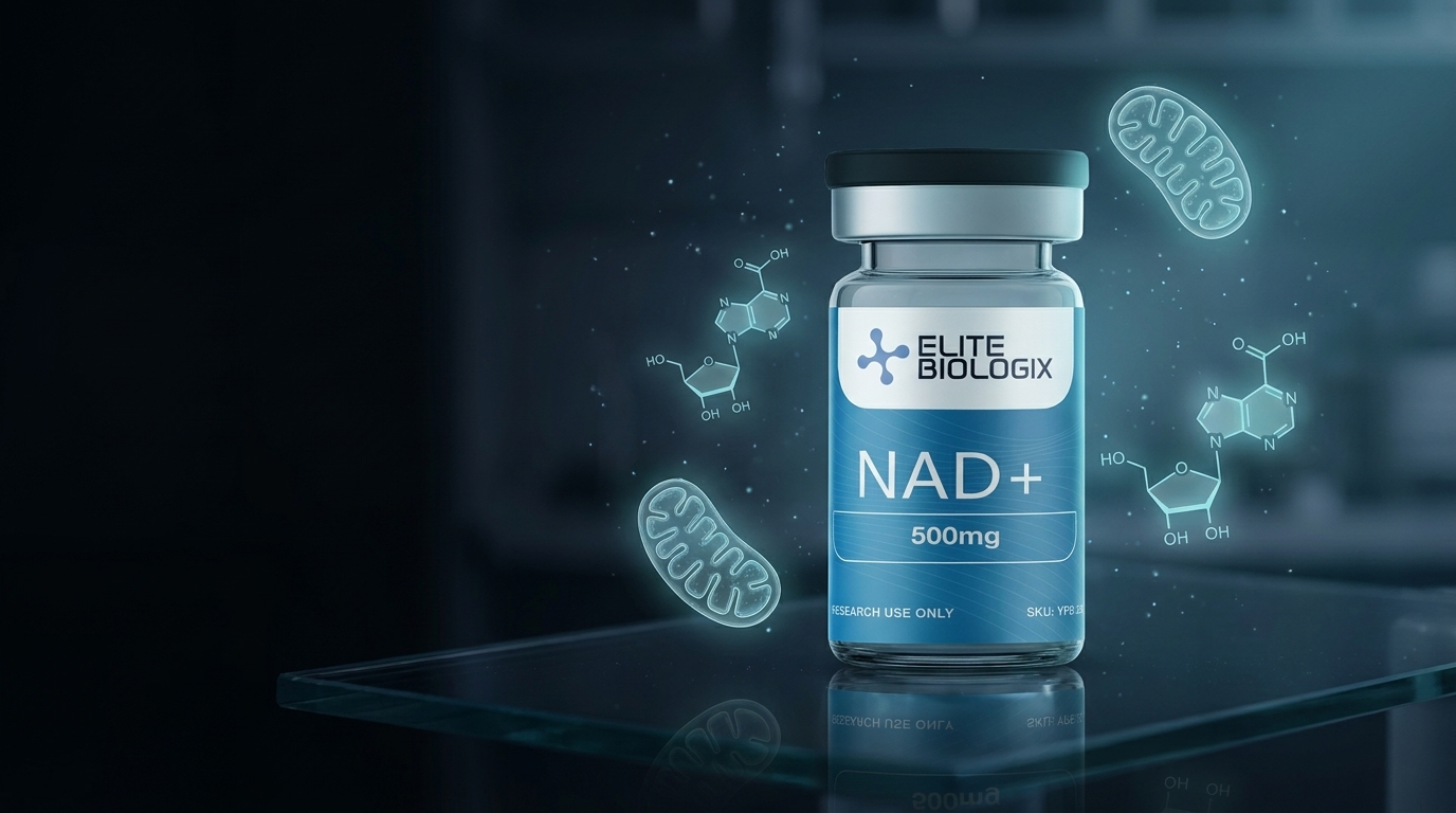
Elite Biologix supplies NAD+ (500 mg, lyophilized) at ≥98% purity, verified by third-party batch testing with a published Certificate of Analysis, specifically for use in qualified research environments. View our NAD+ research compound.
The Sirtuin Pathway: NAD+ as the Rate-Limiting Cosubstrate for Deacylase Activity
Sirtuins (SIRT1–7) are NAD+-dependent protein deacylases and ADP-ribosyltransferases that regulate a broad range of aging-associated processes: mitochondrial biogenesis (SIRT1/PGC-1α), stress resistance (SIRT1/FOXO), inflammatory signaling (SIRT1/NF-κB), and mitochondrial electron transport efficiency (SIRT3/Complex I, III, V). Because sirtuins consume NAD+ stoichiometrically in each deacylation reaction, their activity is directly proportional to intracellular NAD+ concentration — when NAD+ falls with age, sirtuin activity falls proportionally.
The foundational evidence linking age-related NAD+ decline to sirtuin dysfunction came from Gomes and colleagues in Sinclair's laboratory: in 22- and 30-month-old mice, declining nuclear NAD+ caused ATP content to fall, cytochrome c oxidase activity to decline, and mitochondrial-encoded oxidative phosphorylation subunit expression to decrease. Restoring NAD+ via NMN supplementation recovered all these parameters to young-mouse levels — and this recovery was abolished by genetic deletion of SIRT1, confirming that the mitochondrial rescue was SIRT1-dependent (Gomes et al., Cell, 2013, PMID: 24360282).
The connection between PARP-1 and sirtuin competition for NAD+ was established by Bai and colleagues in Auwerx's laboratory. PARP-1 knockout mice showed robustly elevated NAD+ in brown adipose tissue and skeletal muscle, reduced PGC-1α acetylation (indicating strong SIRT1 activation) by 40–90%, and higher mitochondrial content with protection against metabolic disease — all without any exogenous NAD+ supplementation. This established that PARP-1 is a major physiological competitor of sirtuins for the shared NAD+ substrate pool (Bai et al., Cell Metabolism, 2011, PMID: 21459330).
CD38: The Understudied NAD+ Consumer That Drives Age-Related Decline
While the sirtuin and PARP pathways have dominated the NAD+ aging literature, Eduardo Chini's group at the Mayo Clinic has built a compelling preclinical case for CD38 — a multifunctional ectoenzyme — as the primary driver of the age-related NAD+ decline. CD38 hydrolyzes NAD+ to produce cADPR (cyclic ADP-ribose) and ADPR second messengers involved in calcium signaling; unlike sirtuins and PARPs, CD38 can degrade NAD+ to completion without the product inhibition that limits enzymatic activity at very low substrate concentrations.
CD38 expression increases substantially with aging in rodent and human tissues. In 2022, Peclat and colleagues administered the CD38 inhibitor compound 78c to naturally aged mice — not transgenic or disease models, but ordinary aging C57BL/6J mice — and observed a 10% increase in median survival, significantly improved exercise performance, and improved metabolic function (Peclat et al., Aging Cell, 2022, PMID: 35263032). This was the first demonstration that pharmacological CD38 inhibition, in the context of normal aging, could extend lifespan in rodents — making CD38 a potentially important pharmacological target for NAD+ biology researchers.
Research note: The 10% median lifespan extension from CD38 inhibition in naturally aged mice is a mechanistically important finding that receives far less attention in the secondary NAD+ literature than the NMN/NR supplementation studies, despite representing an independent approach to the same biological problem (declining NAD+). CD38 inhibition addresses consumption directly, while precursor supplementation addresses synthesis. Researchers designing NAD+ pathway experiments should consider whether the intervention target is upstream (precursor availability) or downstream (enzymatic consumption), as this distinction affects which endpoints and controls are appropriate.
| NAD+-Consuming Enzyme | Primary Function | Age-Related Change | Key Model Evidence |
|---|---|---|---|
| SIRT1 / SIRT3 | Deacylation of PGC-1α, NF-κB, ETC subunits | Activity falls proportionally with NAD+ decline | PMID 24360282, 21459330 |
| PARP1 | DNA single-strand break repair; PAR chain synthesis | Chronically hyperactivated by accumulated DNA damage; depletes NAD+ | PMID 21459330 |
| CD38 | cADPR/ADPR second messenger production; Ca²⁺ signaling | Expression increases substantially with aging; primary driver of NAD+ depletion | PMID 35263032 |
| All findings from controlled animal model studies. Not validated in human clinical trials for therapeutic applications. | |||
NMN and NR Supplementation in Animal Aging Models
The two best-studied NAD+ precursors in animal models are nicotinamide mononucleotide (NMN) and nicotinamide riboside (NR). Both increase intracellular NAD+ via the NAD+ salvage pathway, though they enter at different enzymatic steps: NMN via the NMNAT enzymes directly, and NR via the NRK1/NRK2 kinases followed by NMNAT.
Yoshino and colleagues in Imai's laboratory demonstrated that NMN supplementation restored compromised NAD+ levels in key metabolic organs in both diet-induced and age-induced diabetic mouse models, significantly improving glucose intolerance, hepatic insulin sensitivity, lipid profiles, and SIRT1-mediated gene expression related to oxidative stress and circadian rhythms (Yoshino et al., Cell Metabolism, 2011, PMID: 21982712). A 12-month long-term study in aging mice found that NMN at 100 or 300 mg/kg/day suppressed age-associated body weight gain (dose-dependently, ~4–9% reduction) and significantly improved insulin sensitivity without adverse effects (Mills et al., Cell Metabolism, 2016, PMID: 28068222).
The most mechanistically significant NR data came from a landmark 2016 Science paper by Zhang, Ryu, Wu, Gariani, and colleagues across both Sinclair's and Auwerx's laboratories: NR supplementation in aged mice rejuvenated muscle stem cells via mitochondrial unfolded protein response (UPRmt) and prohibitin synthesis, delayed senescence in neural stem cells and melanocyte stem cells, and extended mouse lifespan across multiple experimental cohorts — a multi-tissue, multi-cell-type effect driven by NAD+ restoration and sirtuin reactivation (Zhang et al., Science, 2016, PMID: 27127236).
Foundational work from Brenner's laboratory established that NR extends lifespan in yeast via two independent metabolic pathways — the Nrk1 pathway and the Urh1/Pnp1/Meu1 pathway — both converging on NAD+ synthesis, with downstream effects mimicking caloric restriction via Sir2-dependent silencing (Belenky et al., Cell, 2007, PMID: 17482543).
NAD+ in Neurovascular and Neurological Aging Models
The application of NAD+ precursor supplementation to neurovascular aging models represents an active research frontier. Tarantini and colleagues demonstrated that NMN supplementation in aged mice (24 months) over 2 weeks significantly rescued neurovascular coupling responses impaired by aging, improving spatial working memory and gait coordination through increased endothelial NO-mediated vasodilation — a finding that links NAD+/sirtuin biology to cerebrovascular function in aged rodents (Tarantini et al., Innovation in Aging, 2020, PMC: 7741797).
In a neurodegeneration model, Hou and colleagues at the National Institute on Aging demonstrated that NR supplementation in an Alzheimer's disease mouse model (AD/Polβ, combining amyloid pathology with DNA repair deficiency) completely reversed memory-retention deficits in the Morris water maze, dramatically decreased phospho-tau at all measured sites, normalized pro-inflammatory cytokines (IL-1α, TNF-α, MCP-1) to wildtype levels, and significantly decreased the DNA damage marker γH2AX in hippocampus (Hou et al., PNAS, 2018, PMID: 29432159). This multi-pathway rescue — covering memory, tau, neuroinflammation, and DNA repair simultaneously — demonstrates that NAD+ restoration can address multiple hallmarks of neurodegeneration simultaneously in this specific rodent model.
| Study (Year) | Animal Model | Intervention | Primary Outcome | PMID |
|---|---|---|---|---|
| Gomes, Sinclair (2013) | Aged C57BL/6 mice | NMN restoration | Restored OXPHOS subunit expression; SIRT1-dependent mitochondrial rescue | 24360282 |
| Bai, Auwerx (2011) | PARP-1 KO mice | PARP-1 deletion | NAD+ elevation; SIRT1 activation (40–90% PGC-1α deacetylation); metabolic protection | 21459330 |
| Zhang, Sinclair+Auwerx (2016) | Aged mice, multiple stem cell models | NR supplementation | Rejuvenated muscle/neural/melanocyte stem cells; extended lifespan | 27127236 |
| Mills, Imai (2016) | Aging C57BL/6 mice (12-month study) | NMN 100 or 300 mg/kg/day | 4–9% reduction in age-associated weight gain; improved insulin sensitivity | 28068222 |
| Hou et al. NIA (2018) | AD/Polβ mouse model | NR supplementation | Reversed memory deficits; normalized tau, cytokines, DNA damage in hippocampus | 29432159 |
| Peclat, Chini (2022) | Naturally aged C57BL/6J mice | CD38 inhibitor 78c | +10% median survival; improved exercise and metabolic function | 35263032 |
| Belenky, Brenner (2007) | Yeast (S. cerevisiae) | NR supplementation | Extended lifespan via Nrk1 + Urh1/Pnp1/Meu1 pathways; Sir2-dependent silencing | 17482543 |
| All findings from controlled animal model studies or yeast models. Not validated in human clinical trials for therapeutic applications. | ||||
Elite Biologix supplies NAD+ (500 mg, lyophilized) at ≥98% purity, verified by third-party batch testing with a published Certificate of Analysis, specifically for use in qualified research environments. View our NAD+ research compound.
Frequently Asked Questions About NAD+ Preclinical Research
What is NAD+ and why do researchers study it in the context of aging?
Nicotinamide adenine dinucleotide (NAD+) is a dinucleotide coenzyme present in every cell, where it functions as an electron carrier in glycolysis, the TCA cycle, and oxidative phosphorylation, and as a required substrate for NAD+-consuming enzymes including sirtuins (SIRT1-7), poly(ADP-ribose) polymerases (PARPs), and CD38. NAD+ levels decline substantially in aged animal tissues — 55–67% in rodent skeletal muscle — and this decline is associated with reduced sirtuin activity, mitochondrial dysfunction, increased inflammation, and impaired DNA repair. Researchers study NAD+ in aging biology because restoring NAD+ levels in animal models has been shown to reverse multiple hallmarks of cellular aging simultaneously.
What is the difference between NMN and NR as NAD+ precursors in animal models?
NMN (nicotinamide mononucleotide) and NR (nicotinamide riboside) are both salvage pathway precursors that increase intracellular NAD+. NR requires the NRK1/NRK2 kinases to convert it to NMN, which then enters NAD+ synthesis via NMNAT enzymes. NMN enters at the NMNAT step directly. Both have demonstrated NAD+-elevating effects in rodent tissues. NMN has more extensive published data in aging-focused mouse models (Imai/Sinclair groups), while NR has demonstrated stem cell rejuvenation and lifespan extension effects (Auwerx/Sinclair 2016 Science paper, PMID 27127236). The primary mechanistic difference in rodent studies is tissue distribution kinetics — ongoing research areas, not settled questions.
What is CD38 and why is it relevant to NAD+ research?
CD38 is a multifunctional ectoenzyme that hydrolyzes NAD+ to produce cADPR and ADPR calcium-signaling second messengers. CD38 expression increases substantially with aging in rodent and human tissues, and its activity can dominate the NAD+ degradation profile in aged cells — consuming NAD+ faster than salvage synthesis can replenish it. The Chini group at Mayo Clinic has established CD38 as a pharmacological target in aging biology: compound 78c, a CD38 inhibitor, extended median survival by 10% in naturally aged (non-transgenic) mice, demonstrating that addressing NAD+ consumption directly has lifespan consequences in normal aging rodent models (PMID: 35263032).
What is the PARP-NAD+ connection in aging?
PARP1 and PARP2 are DNA repair enzymes that consume NAD+ stoichiometrically when synthesizing poly(ADP-ribose) chains at DNA strand break sites. During aging, accumulated DNA damage chronically hyperactivates PARP1, which depletes the NAD+ pool and starves sirtuins of their cofactor — creating a feed-forward degradation loop. PARP-1 knockout mice showed robustly elevated NAD+ content, strong SIRT1 activation (40–90% reduction in PGC-1α acetylation), higher mitochondrial content, and protection against metabolic disease (PMID: 21459330). This established the PARP-sirtuin competition for NAD+ as a major mechanism of age-related metabolic decline in rodent models.
Is NAD+ approved for any therapeutic use in humans?
NAD+ itself is not approved by the FDA or any regulatory authority for the treatment of any disease or condition. Its precursors NMN and NR are available as dietary supplements but have not completed the clinical trial process required for drug approval. All mechanistic and efficacy data cited in this article derives from in vitro assays, yeast models, and controlled rodent studies. NAD+ is sold exclusively for laboratory and research purposes.
Conclusion: NAD+ in Aging Biology Research
The NAD+ preclinical literature is distinguished by the breadth of its mechanistic connections — from electron transport and ATP production to epigenetic regulation, inflammatory signaling, DNA repair, and stem cell maintenance. No other single coenzyme has been demonstrated to influence this many aging-associated biological processes simultaneously in animal models. The convergence of independent research groups (Sinclair, Auwerx, Imai, Brenner, Chini) on overlapping findings across different interventions (NMN, NR, PARP inhibition, CD38 inhibition) represents an unusually high degree of cross-institutional corroboration for a preclinical biology field.
The limitations of this literature are equally important to understand. Rodent aging biology does not reliably translate to human aging biology — differences in NAD+ metabolism, lifespan biology, and disease mechanisms between mice and humans are substantial. The absence of large, controlled human clinical trials means that the mechanistic insights from animal models cannot be applied therapeutically without further validation. Researchers designing NAD+ experiments should carefully consider which intervention targets the consumption side (CD38 inhibition, PARP1 inhibition) versus the synthesis side (NMN, NR precursor supplementation), as the McReynolds 2020 isotope tracer data suggests consumption may be the primary driver of the age-related decline. [INTERNAL-LINK: SS-31 research article → complementary mitochondrial research compound targeting cristae structure and ETC supercomplex architecture] [INTERNAL-LINK: MOTS-c research article → mitochondrially-derived peptide acting on nuclear AMPK and aging pathways]
Elite Biologix supplies NAD+ (500 mg, lyophilized) at ≥98% purity, verified by third-party batch testing with a published Certificate of Analysis, specifically for use in qualified research environments. View our NAD+ research compound.
References
- McReynolds MR, Chellappa K, Baur JA. Age-related NAD+ decline. Experimental Gerontology. 2020. PMID: 32097708.
- Gomes AP, Price NL, Ling AJY, Moslehi JJ, Montgomery MK, Rajman L, White JP, Teodoro JS, Wrann CD, Hubbard BP, Mercken EM, Palmeira CM, de Cabo R, Rolo AP, Turner N, Bell EL, Sinclair DA. Declining NAD(+) induces a pseudohypoxic state disrupting nuclear-mitochondrial communication during aging. Cell. 2013;155(7):1624–38. PMID: 24360282.
- Bai P, Cantó C, Oudart H, Brunyánszki A, Cen Y, Thomas C, Yamamoto H, Huber A, Kiss B, Houtkooper RH, Schoonjans K, Schreiber V, Sauve AA, Menissier-de Murcia J, Auwerx J. PARP-1 inhibition increases mitochondrial metabolism through SIRT1 activation. Cell Metabolism. 2011;13(4):461–8. PMID: 21459330.
- Zhang H, Ryu D, Wu Y, Gariani K, Wang X, Luan P, D'Amico D, Ropelle ER, Lutolf MP, Aebersold R, Schoonjans K, Menzies KJ, Auwerx J. NAD+ repletion improves mitochondrial and stem cell function and enhances life span in mice. Science. 2016;352(6292):1436–43. PMID: 27127236.
- Mills KF, Yoshino S, Stein LR, Grozio A, Kubota S, Sasaki Y, Redpath P, Migaud ME, Apte RS, Uchida K, Yoshino J, Imai SI. Long-term administration of nicotinamide mononucleotide mitigates age-associated physiological decline in mice. Cell Metabolism. 2016;24(6):795–806. PMID: 28068222.
- Yoshino J, Mills KF, Yoon MJ, Imai S. Nicotinamide mononucleotide, a key NAD(+) intermediate, treats the pathophysiology of diet- and age-induced diabetes in mice. Cell Metabolism. 2011;14(4):528–36. PMID: 21982712.
- Peclat TR, Thompson KL, Warner GM, Chandra LC, Witte MA, Mazer-Amirshahi M, Tarantini S, Csiszar A, Ungvari Z, Johnson SD, Chini CCS, Chini EN. CD38 inhibitor 78c protects against aging-associated metabolic dysfunction and extends healthspan. Aging Cell. 2022. PMID: 35263032.
- Hou Y, Lautrup S, Cordonnier S, Wang Y, Croteau DL, Zavala E, Zhang Y, Moritoh K, O'Connell JF, Baptiste BA, Stevnsner TV, Mattson MP, Bohr VA. NAD+ supplementation normalizes key Alzheimer's features and DNA damage responses in a new AD mouse model with introduced DNA repair deficiency. PNAS. 2018;115(8):E1876–E1885. PMID: 29432159.
- Belenky P, Racette FG, Bogan KL, McClure JM, Smith JS, Brenner C. Nicotinamide riboside promotes Sir2 silencing and extends lifespan via Nrk and Urh1/Pnp1/Meu1 pathways to NAD+. Cell. 2007;129(3):473–84. PMID: 17482543.
- Tarantini S, Valcarcel-Ares MN, Toth P, Yabluchanskiy A, Tucsek Z, Kiss T, Hertelendy P, Kinter M, Ballabh P, Süle Z, Farkas E, Baur JA, Sinclair DA, Csiszar A, Ungvari Z. Nicotinamide mononucleotide (NMN) supplementation rescues cerebromicrovascular endothelial function and neurovascular coupling responses and improves cognitive function in aged mice. Redox Biology. 2019;24:101192. PMC: 7741797.
NAD+ is sold exclusively for laboratory and research purposes. It is not approved for human use by the FDA or any regulatory authority. All information presented in this article is derived from published preclinical research using in vitro assays, yeast models, and animal models. Findings from these models cannot be presumed to translate directly to human physiology or to constitute clinical evidence of therapeutic efficacy. Elite Biologix does not make any claims regarding the safety or efficacy of NAD+ for use in humans or animals.